The human foot is one of the most complex and specialized structures in the body, composed of 26 bones, 33 joints, and more than 100 muscles, tendons, and ligaments that work in perfect coordination.
This complex architecture has evolved over millions of years to enable efficient bipedal locomotion, becoming a masterpiece of biomechanical engineering.
Understanding how gait biomechanics works is not just an academic exercise, but essential knowledge for anyone who walks or runs regularly.
Proper footwork directly influences movement efficiency, injury prevention, and athletic performance. Every step we take involves a precise sequence of biomechanical events that are repeated thousands of times during normal activity.
Problems with gait biomechanics can have consequences that extend far beyond the foot, affecting the ankles, knees, hips, and even the spine.
For this reason, understanding the fundamental principles of how the foot works during walking and running is essential to maintaining an active and healthy life.
Functional anatomy of the foot
To understand the biomechanics of gait, it is necessary to first understand the anatomical structure of the foot and how each component contributes to its function during movement.
Bone and joint structure
The foot is anatomically divided into three main regions: the hindfoot, the midfoot, and the forefoot. The hindfoot includes the calcaneus and talus, bones that form the subtalar joint, crucial for inversion and eversion movements. This joint allows the foot to adapt to uneven surfaces and absorb impact forces.
The midfoot comprises the five tarsal bones, including the navicular, cuboid, and three cuneiforms. These bones form the medial longitudinal arch, a critical structure for shock absorption and propulsion. The integrity of this region is crucial for normal foot function.
The forefoot includes the five metatarsals and the phalanges of the toes. The metatarsals act as levers during toe-off, while the toes provide stability and contribute to final propulsion during walking and running.
Ligamentous and fascial system
The plantar fascia is a thick fibrous structure that extends from the calcaneus to the metatarsal heads, acting as a tension cable that maintains the longitudinal arch. During walking, this structure stretches and contracts, storing and releasing elastic energy that significantly contributes to movement efficiency.
The plantar ligaments provide passive stability to the multiple joints of the foot, while the lateral and medial ankle ligaments control movements in the frontal plane. The integrity of these structures is critical for maintaining stability during dynamic activities.
Intrinsic and extrinsic muscles
The intrinsic muscles of the foot, located entirely within the foot structure, provide fine control and stabilization of the metatarsophalangeal joints. These muscles include the interossei, lumbricals, and the muscles of the great toe, which work constantly to maintain stability and control during movement.
Strengthening and properly activating these intrinsic muscles is critical for optimal biomechanics. Products specifically designed to stimulate and support these muscles, such as biomechanical socks with graduated compression and arch support , can help improve the function of these muscles during physical activity.
The extrinsic muscles, which originate in the leg and insert into the foot, provide the primary driving force for movement. The triceps surae (gastrocnemius and soleus) are essential for propulsion, while the anterior and posterior muscles of the leg control dorsiflexion and plantarflexion.

Phases of normal walking
The gait cycle is divided into specific phases, each with distinct biomechanical characteristics that require precise coordination between all components of the foot and lower limb.
Initial contact phase
Initial contact typically occurs on the posterior-lateral aspect of the heel, although this pattern can vary depending on walking speed and individual characteristics. At this point, the foot must absorb impact forces while it begins to adapt to the terrain.
During this phase, the subtalar joint initiates a controlled eversion movement that allows the foot to become more flexible and adapt to the surface. This movement is coordinated with the internal rotation of the tibia, creating an efficient shock absorption mechanism.
Medium support phase
During midstance, the entire foot is in contact with the ground and must function as a stable platform to support the body's weight. The subtalar joint reaches its maximum eversion, and the longitudinal arch experiences its greatest deformation.
During this phase, the intrinsic muscles of the foot work intensively to maintain stability, while the plantar fascia stretches to absorb energy. Foot function during this phase is critical for maintaining balance and preparing for propulsion.
Propulsion phase
The propulsion phase begins when the heel lifts off the ground and culminates with toe-off. During this phase, the subtalar joint rapidly reverses, converting the foot into an efficient, rigid lever for propulsion.
The windlass mechanism is activated when the toes extend, tensing the plantar fascia and elevating the arch. This mechanism transforms the elastic energy stored during stance into propulsive force, significantly contributing to the energy efficiency of gait.
During running, impact forces can reach 2.5 to 3 times body weight.
Biomechanical differences between walking and running
Although the basic principles of foot biomechanics are similar in walking and running, there are important differences in the forces, timing, and patterns of movement.
Magnitude of forces
During normal walking, impact forces typically reach 1.2–1.5 times body weight. In contrast, during running, these forces can reach 2.5–3 times body weight, requiring specific adaptations in foot structure and function.
These increased forces during running require greater impact absorption and stabilization capacity, which explains why certain biomechanical problems that may be asymptomatic during walking become apparent during running.
Temporal patterns
The gait cycle includes a double support phase, where both feet are in contact with the ground simultaneously. This phase disappears during running, being replaced by a flight phase, where neither foot is in contact with the ground.
The absence of a double support phase during running means that each foot must provide all of the stability and control during its stance phase, increasing the demands on the neuromuscular control systems.
Neuromuscular adaptations
Running requires greater activation of the stabilizing muscles of the foot and ankle due to the increased forces and the absence of a double-support phase. The intrinsic muscles of the foot must work harder to maintain stability during more dynamic landings.
The temporal coordination between different muscle groups also changes during running, with more pronounced anticipatory activation to prepare the foot for the increased impact.

Pronation and supination: normal vs. pathological movements
Pronation and supination movements are normal and essential components of foot biomechanics, but they can become problematic when they occur excessively or at inappropriate times in the gait cycle.
Normal pronation
Pronation is a triplanar movement that combines eversion of the subtalar joint, abduction, and dorsiflexion. This movement is essential for impact absorption and adaptation to the terrain during the initial phases of ground contact.
Normal pronation typically reaches its peak around 40–60% of the stance phase, followed by rapid resupination that prepares the foot for propulsion. This precise timing is crucial for efficient biomechanical function.
Overpronation
Overpronation can manifest as excessive range of motion, inappropriate timing, or inadequate pronation velocity. Excessive overpronation can overload the medial structures of the foot and alter the biomechanics of the entire lower extremity.
Runners with overpronation may experience increased stress on the plantar fascia, posterior tibial tendon, and medial knee structures. Identifying and managing overpronation is important for preventing overuse injuries.
Supination and rigid foot
Supination is the opposite movement to pronation, combining subtalar inversion with adduction and plantar flexion. While supination is normal during propulsion, excessive or persistent supination can limit the foot's shock absorption capacity.
Supinated or rigid feet may transmit more impact to proximal structures and may be at greater risk for stress injuries to the bones of the foot and leg.
The windlass mechanism can contribute up to 17% of the total energy required for locomotion.
The plantar arch: structure and function
The plantar arch is one of the most distinctive features of human anatomy and plays a fundamental role in the biomechanical function of the foot during locomotion.
Anatomy of the longitudinal arch
The medial longitudinal arch is the most prominent structure, formed by the calcaneus, talus, navicularis, cuneiform bones, and the first three metatarsals. This structure is actively supported by muscles and passively supported by ligaments and the plantar fascia.
The lateral longitudinal arch, although less prominent, also contributes to foot function. It is formed by the calcaneus, cuboid, and the fourth and fifth metatarsals, providing lateral stability during movement.
Energy absorption function
During the loading phase, the arch partially flattens, storing elastic energy in the plantar fascia and plantar ligaments. This controlled deformation acts as a natural shock absorption mechanism that reduces forces transmitted to proximal structures.
The amount of arch deformation is finely regulated by muscular activity, especially the intrinsic muscles of the foot and the posterior tibialis. This active regulation allows adaptation to different surfaces and loading conditions.
Energy return mechanism
During propulsion, the arch rapidly returns to its original configuration, releasing stored elastic energy and contributing to the energy efficiency of movement. This energy return mechanism can contribute up to 17% of the total energy required for locomotion.
The timing of this energy return mechanism is coordinated with the activation of the windlass mechanism, creating an efficient, integrated propulsion system that characterizes human locomotion.

Windlass and propulsion mechanism
The windlass mechanism is a crucial component of the propulsive biomechanics of the foot that transforms the foot from a flexible structure to a rigid lever during takeoff.
Activation of the mechanism
The mechanism is activated when the metatarsophalangeal joints extend during the propulsive phase of gait. This extension tenses the plantar fascia, which acts like a cable connecting the heel to the toes.
The tension of the plantar fascia elevates the longitudinal arch and transforms the foot into a rigid structure that can efficiently transfer the muscular force of the triceps surae to the ground during propulsion.
Coordination with other systems
The windlass mechanism is temporally coordinated with the inversion of the subtalar joint and the activation of the triceps surae. This coordination creates an integrated propulsive system that maximizes energy efficiency.
Dysfunction in any of these components can compromise the efficiency of the windlass mechanism, resulting in increased energy expenditure and potential overload of other structures.
Factors affecting function
The stiffness of the plantar fascia, the mobility of the metatarsophalangeal joints, and the strength of the intrinsic muscles of the foot influence the efficiency of the windlass mechanism. Limitations in any of these factors can compromise propulsive function.
Age, activity level, and individual anatomical characteristics also affect the function of the windlass mechanism, explaining some of the individual variability in gait efficiency and susceptibility to injury.
Adaptations of the foot to different surfaces
The foot must constantly adapt to changing terrain characteristics, a process that requires complex, real-time adjustments of multiple biomechanical systems.
Hard vs. soft surfaces
On hard surfaces like asphalt or concrete, the foot must absorb more impact due to the lack of surface deformation. This requires greater muscle activation and can result in greater fatigue of the stabilizing muscles.
On soft surfaces like sand or grass, the foot must provide greater active stability due to the surface's inherent instability. This can result in greater intrinsic muscle activation and greater energy expenditure.
Uneven terrain
Uneven terrain requires constant adjustments in foot position and stiffness to maintain stability and prevent injury. These adjustments are mediated by the proprioceptive system and require rapid reaction time.
Regular exposure to uneven terrain can improve proprioceptive function and stabilizing muscle strength, but can also increase the risk of acute injury if adaptation is inadequate.
Inclines and slopes
Walking or running on slopes requires specific adaptations in foot biomechanics. On upward slopes, the ankle must maintain greater dorsiflexion, while on downward slopes, it must control excessive plantar flexion.
These adaptations can overload different muscle groups and structures in the foot, explaining why certain biomechanical problems manifest specifically during activities on inclined terrain.

Factors influencing individual biomechanics
Foot biomechanics varies significantly between individuals due to multiple anatomical, physiological, and environmental factors that interact in complex ways.
Anatomical factors
The bone structure of the foot, including arch height, metatarsal length, and calcaneus shape, significantly influences biomechanical patterns.
These anatomical variations are largely genetically determined and may predispose to certain movement patterns.
Joint flexibility and ligament laxity also vary considerably between individuals, affecting the range and execution of movements during the gait cycle. These characteristics can change with age, training, and previous injuries.
Neuromuscular characteristics
The strength, endurance, and coordination of the foot and leg muscles vary among individuals and can be modified through specific training. These neuromuscular characteristics directly influence the ability to control and stabilize movement.
Neuromuscular activation patterns can also vary, with some people showing greater reliance on passive versus active control strategies. These differences can influence susceptibility to certain types of injuries.
External factors
Footwear has a significant impact on foot biomechanics, altering impact forces, movement patterns, and muscle activation. Different types of footwear can substantially alter the natural biomechanics of the foot.
The training surface used can also influence long-term biomechanical adaptations. Runners who train predominantly on hard surfaces may develop different adaptations than those who train on a variety of surfaces.
Foot biomechanics in different populations
The biomechanical characteristics of the foot vary according to age, sex, activity level, and other population characteristics, requiring specific considerations for each group.
Age differences
In children, the biomechanics of the foot is constantly developing, with gait patterns gradually maturing until adolescence. The arches of the foot develop progressively, and neuromuscular patterns are refined with motor experience.
In older adults, age-related changes may include loss of flexibility, decreased muscle strength, and changes in proprioception. These changes may require adaptations to footwear and training strategies.
Differences between sexes
Women tend to exhibit greater joint flexibility and different muscle activation patterns compared to men. These differences may influence susceptibility to certain types of injuries and may require specific considerations in footwear design.
Hormonal differences can also affect foot biomechanics, especially during periods of significant hormonal changes such as pregnancy or menopause.
Athletes vs. general population
Athletes often develop specific biomechanical adaptations related to their sport, which may include optimized movement patterns but also increased susceptibility to overuse injuries.
The specific demands of different sports may result in particular biomechanical adaptations that may not be appropriate for activities of daily living or other sports.

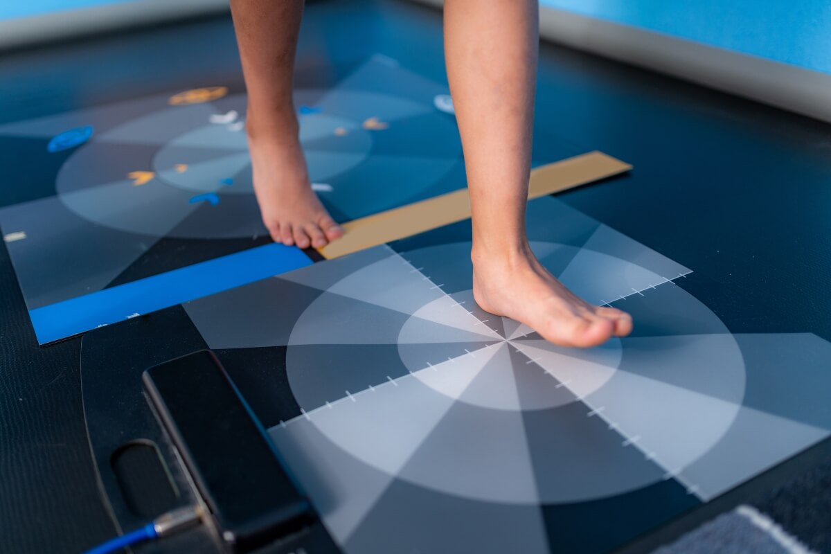
Evaluation of the biomechanics of the tread
The assessment of gait biomechanics can be performed at different levels of complexity, from simple clinical observation to sophisticated laboratory analysis.
Clinical observation
Visual gait observation may reveal obvious asymmetries, abnormal shoe wear patterns, and obvious compensations. This assessment, although limited, can identify significant biomechanical problems that require attention.
Footwear wear assessment provides valuable information on long-term loading patterns and can reveal asymmetries or abnormal patterns that are not evident during direct observation.
Instrumental analysis
Video-recorded gait analysis allows for a more detailed assessment of movement patterns, timing, and coordination. These tools can quantify specific parameters and provide objective information on biomechanical function.
Force platforms can measure ground reaction forces and provide information on loading patterns during different phases of the gait cycle. This information is valuable for understanding the mechanical demands on different structures.
Advanced technology
Three-dimensional gait analysis systems can provide detailed information on the kinematics and kinetics of the entire lower limb. These systems are especially useful for the investigation and evaluation of complex cases.
Electromyography can assess muscle activation patterns during movement, providing information about neuromuscular function that is not visible through other assessment methods.
Clinical and therapeutic implications
Understanding the biomechanics of gait has important implications for the treatment and prevention of foot and lower extremity injuries.
Identification of risk factors
Abnormal biomechanical patterns can predispose to specific injuries. Early identification of these patterns allows for preventative interventions that can significantly reduce injury risk.
Biomechanical assessment can also explain why certain injuries recur or do not respond to conventional treatment, guiding more targeted and effective therapeutic strategies.
Intervention strategies
Biomechanical interventions may include specific exercises to strengthen weak muscles, stretches to improve limited flexibility, and neuromuscular reeducation techniques to correct abnormal movement patterns.
Therapeutic footwear and orthotic devices can modify foot biomechanics when exercise interventions are not sufficient or when more significant modifications are needed.
In this context, biomechanical socks represent an innovation that combines comfort with therapeutic functionality, providing targeted support to the intrinsic muscles of the foot and optimizing the function of the plantar arch during movement.
Progress monitoring
Repeated biomechanical assessment can monitor the effectiveness of interventions and guide adjustments to the treatment plan. This objective assessment is especially important in complex cases or when progress is slow.

Emerging technologies in foot biomechanics
Technological advances are revolutionizing our ability to understand and evaluate foot biomechanics, opening up new possibilities for diagnosis and treatment.
Innovations in biomechanical textiles
The development of technical textiles has enabled the creation of products that not only provide comfort but also actively influence foot biomechanics.
Podoks biomechanical socks represent a significant evolution in this field, incorporating graduated compression technologies that can stimulate proprioception, support the plantar arch, and optimize intrinsic muscle activation.
These products utilize specific compression patterns designed by podiatric professionals to work in synergy with the natural anatomy of the foot, providing support where needed without interfering with normal biomechanical function.
Portable sensors
Wearable sensors can provide real-time biomechanical information during daily activities, beyond laboratory assessments. This technology allows for a better understanding of how the foot functions under real-life conditions.
These devices can monitor parameters such as plantar pressure, joint angles, and muscle activation patterns over extended periods, providing information that cannot be obtained through single-spot assessments.
Computational modeling
Advanced computational models can simulate foot biomechanics under different conditions, allowing predictions about the effects of specific interventions without the need to physically implement them.
These models can assist in the design of customized footwear, optimized orthotic devices, and more effective rehabilitation strategies.
Artificial intelligence
Artificial intelligence algorithms can analyze large amounts of biomechanical data to identify subtle patterns that might not be apparent to traditional human analysis.
This technology can help in the early identification of risk factors, the personalization of interventions, and the prediction of therapeutic outcomes.
Conclusions
Foot biomechanics represents a complex and integrated system that has evolved to enable efficient and adaptable locomotion. Understanding the fundamental principles of how the foot functions during walking and running is essential for anyone involved in activities requiring locomotion, from recreational walkers to elite athletes.
Individual variability in foot biomechanics is considerable and is influenced by multiple anatomical, physiological, and environmental factors. This variability means that there is no universally "perfect" biomechanical pattern; rather, each individual must be evaluated and treated according to their specific characteristics.
Advances in assessment technology and our growing understanding of foot biomechanics are opening up new possibilities for the prevention and treatment of problems related to foot function. However, it is important to remember that biomechanics is only one component of the overall function of the musculoskeletal system and must be considered within the broader context of an individual's overall health and well-being.
-----
Scientific references:
Perry, J., & Burnfield, J.M. (2010). "Gait Analysis: Normal and Pathological Function." SLACK Incorporated, 2nd Edition.
Cavanagh, P.R., & Lafortune, M.A. (1980). "Ground reaction forces in distance running." Journal of Biomechanics, 13(5), 397-406.
Ker, R.F., et al. (1987). "The spring in the arch of the human foot." Nature, 325(7000), 147-149.
Hicks, J. H. (1954). "The mechanics of the foot: II. The plantar aponeurosis and the arch." Journal of Anatomy, 88(1), 25-30.
Nigg, B.M., et al. (2015). "Running shoes and running injuries: mythbusting and a proposal for two new paradigms." British Journal of Sports Medicine, 49(20), 1290-1294.
McKeon, P.O., et al. (2015). "The foot core system: a new paradigm for understanding intrinsic foot muscle function." British Journal of Sports Medicine, 49(5), 290.
Daoud, A.I., et al. (2012). "Foot strike and injury rates in endurance runners: a retrospective study." Medicine & Science in Sports & Exercise, 44(7), 1325-1334.


Leave a comment
All comments are moderated before being published.
This site is protected by hCaptcha and the hCaptcha Privacy Policy and Terms of Service apply.